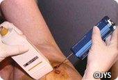
 When the clinician or radiologist can feel a lesion or identifies one from a mammography or during a breast ultrasound then in order to confirm whether it is benign or malignant the professional needs to analyse some of the cells.
- When the suspicious area is a cyst then a fine needle aspiration is commonly performed to withdraw a sample of the liquid for further analysis. At the same time all the liquid from the cyst can be withdrawn though this is not a definitive treatment plan for cysts.
- When the suspicious area is solid, or a dense zone, or made up of micro-calcifications or indeed for any lesion requiring further investigation then a biopsy is required to analyse the tissue. The biopsy entails removal of a small area of the suspicious region which will be examined under the microscope (this is a histological examination or an anatomopathological examination). Several biopsies types exist: micro biopsy, macro biopsy, single intact biopsy system and surgical (open) biopsy
Histological examination of the tissue is the only way to identify the exact cancerous nature of any lesion. For the examination to be conducted properly, collaboration between the radiologist, the clinician and the anatomopathologist is key. This is why a top quality experienced team that you can rely on is so important when attempting to treat this type of pathology.


