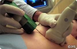
An abnormality is identified most often with ultrasound. Then a small cylinder (or carrot) of the identified tissue is removed.
A local anesthetic is given and a type of automatic gun is guided via the ultrasound to the abnormality and small fragments of the tissue are removed.

Because a local anesthetic is given, this biopsy is almost painless.
The histological results are interpreted by an anatomopathologist and returned in under a week and most often within 3 days. Therefore this allows to be quickly reassured if the lesion is benign or if the lesion is malignant, suggest the best treatment quickly.


