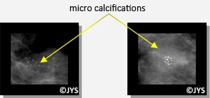
A mammogram is an x-ray image of both breasts which looks for:
- 1-variations in breast tissue density, where the breast is compressed between two plates to flatten the area and better locate any abnormalities.

- 2- calcium deposits often in the form of calcified dust particles (called micro-calcifications) some of which require verification as to their importance.

Mammograms require only low dose radiation (and do not impact the risk of breast cancer). They can even be performed if necessary during pregnancy, once certain safety precautions are respected.
Some more modern techniques now available, such as the digital mammography, enable improved image quality which is an extra advantage in the case of younger women who have very dense breasts.
The breast ultrasound is useful when associated with a mammogram in order to facilitate a more precise image, or perform a guided biopsy or tissue withdrawal.
Mammograms do not diagnose breast cancer. They detect clinical abnormalities which are then classified according to their probability of being cancerous.
For certain types of cancer, mammograms can return false normal results. It is therefore advisable that mammograms be repeated every 2 years and that they be conducted with a breast ultrasound.


