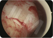
The technique used to remove one or several myomas (fibroids) depends on the number of myomas to be removed, their size and their location.
Myomectomy techniques that remove myomas whilst preserving the uterus and the possibility of future pregnancy under healthy conditions.
Before performing any myomectomy procedure, extensive pre-operative imaging (called mapping) is conducted to identify the exact location of the myoma(s) and measure it/them in order to recommend the most appropriate surgical myomectomy procedure.
Pre-operative imaging assessment includes:
- pelvic ultrasound scans (via the vagina for best results) that map each myoma
- MRI (magnetic resonance imaging) scans of the pelvis
- pre-operative diagnostic hysteroscopy (for situations where the myoma(s) is/are completely or partially submucosal (ie intra cavitary)).
These tests allow the surgeon to identify the location of the myoma(s) vis-a-vis the uterine cavity and decide on the best treatment technique.
Three types of myomas in terms of their position vis-a-vis the uterine cavity:
submucosal; inside the uterus on the mucous membrane or endometrium layer,
insterstital (intramural); within the muscle wall of the uterus
subserosal; on the outside of the uterus wall
Each type requires a specific treatment technique.
Myomectomy via operative hysteroscopy (via the body’s natural pathways/vagina):
When fibromas grow on the inner surface of the uterine cavity they are termed submucosal fibroids.
Submucosal fibromas can be removed with an operative hysteroscopy procedure.
As with other operative hysteroscopy treatments, a light general anesthesia is administered and after a few hours of monitoring, the patient can go home on the day of the procedure.
An operative hysteroscopy is inserted into the uterine cavity and it is equipped with a resectoscope (a cutting loop). The loop cuts the fibroma(s) into thin slices (as a plane might slice a potato into thin strips).

The slices of fibroma (myoma) are extracted from the uterus and then sent for analysis (to verify their constitution).
After the operation the patient suffers no pain but may experience some light bleeding.
Operative hysteroscopy has its limits and cannot remove all fibromas (myomas). Conditions for successful operative hysteroscopy:
- fibromas (myomas) should be less than 40 – 50 mm in size
- the uterine wall supporting the fibromas should be greater than 5 mm thick
- the fibroma should not have other fibromas adjacent to it as this makes the procedure difficult to conduct with uncertain results
A thorough pre-operative assessment is essential for successful treatment. Key here is an ultrasound scan of the vagina (also called mapping) and a diagnostic hysteroscopy.
For situations requiring further examination of the uterine lesions pre-operative tests may also include an MRI (magnetic resonance imaging).
Myomectomy via laparotomy:
One or more myomas are removed via an incision in the abdomen. This is often a horizontal incision made at the bikini like (just at the level of the pubic hair as is often made for a caesarean section delivery) called a Pfannenstiel incision.
Laparotomy procedures are more likely used when several myomas are detected or when they are large.
An incision is made through the uterine wall that exposes the myoma(s) and they can then be removed. This surgery requires relatively significant incisions and can be a cause of haemorrhage.
When the myoma(s) are removed the incision(s) are closed and stitched so as to render the uterine wall as strong as possible. Nevertheless the uterus will always be slightly weaker (contains scar tissue) and requires close monitoring during any subsequent pregnancies.
Myomectomy via laparoscopy:
This technique removes one or more (though not a great number) using an endoscope equipped with a visual aid. Entry to the abdomen is via an incision close to the belly button (navel) and via 3 incisions of 1 cm in length at the base of the abdomen.
This technique is used for single myoma or when there are only a few small myomas to be removed. The time spent with this procedure is more or less identical to a laparotomy, however after myoma removal, the time spent stitching is less as it is often just carried out at one single level.
Myomectomy via the vaginal pathway:
This technique removes subserosal and interstitial myomas via an incision high up in the vagina. This technique is used when there are only a few myomas and when they are accessible via the vagina.


