What are Fibromas or Myomas?
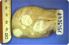
Fibromas are benign tumors. Doctors also term them fibroids, fibroid tumors, myomas, fibromyomas, or leiomyomas. They are the result of a proliferation of muscle cells arising from the uterine wall. They vary in size from pea sized to tennis ball size to even, in rare cases, grapefruit size or even bigger. These tumors have the consistency of a golf ball.


Fibromas resemble balls (myomas) which grow on a wall (the lining of the uterus).
-
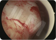
 When fibromas occur within the uterine cavity they are termed submucosal fibromas. They can provoke heavy or prolonged bleeding (menorrhagia) sometimes with clots. They can also lead to infertility since the ball like growth can inhibit successful embryo implantation.
-
 When fibromas grow on the outside of the uterus they are termed subserosal fibromas. Unless they are very large, these rarely cause any symptoms. In terms of size, large fibromas can measure several tens of centimeters resembling a melon or bigger. In these cases such fibromas can compromise the bladder, the urethras, and leave the patient with the feeling of wanting to pass urine to the extent that the kidneys may even dilate. There may also be difficulties in giving birth when the baby is sitting low (Praevia).
-
 When fibromas grow inside the uterine wall (between the uterine cavity and the outside of the uterus) they are terms interstitial fibromas. This is considered as an intermediate situation and does not always exhibit any symptoms. These fibromas are not a problem as such but they can require attention when they become large and voluminous and when they risk inhibiting fertility (especially as regards IVF (in vitro fertilisation) treatments).
In reality, multiple fibromas can be a mix of these three cases and require specific tailored treatment based on the results of ultrasound scans, hysteroscopy procedures and sometimes MRI (magnetic resonance imaging) scans.
Fibromas or myomas are benign tumors. Nevertheless caution is required to verify the absence of any associated lesions which may themselves be malignant especially for post-menopausal patients.
Myomas are benign tumors in the uterus made up of muscular tissue and they can lead to pain, bleeding or problems with falling pregnant. When myomas have no symptoms then they are monitored and do not need to be removed or treated.
How are submucosal fibromas removed?
When fibromas occur within the uterine cavity they are termed submucosal fibromas. These fibromas can be removed with operative hysteroscopy.
How are submucosal fibromas removed?
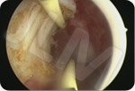
As with all operative hysteroscopy procedures, removal of submucosal fibromas is carried out with a low dose of general anesthesia and the patient is free to return home on the day of the operation. An operating hysteroscope complete with a resection loop is inserted into the uterine cavity. The loop is to cut the fibroma into thin layers (much like a greater slices a potatoe into thin strips). At the end of the removal procedure, each of the layers is removed and analysed.
Some light bleeding but no pain is experienced after this procedure.

Unfortunately however not all submucosal fibroma can be removed using the operative hysteroscopy procedure. This is due to:
- The size of the fibroma – optimally they should be smaller than 40 – 50 millimeters,
- The position of the fibroma – the wall from which they grow should be at least 5 millimeters deep,
- The number of fibromas – these fibromas should not be linked with other adjacent myomas which can lead to unclear procedure results
These considerations indicate the importance of a complete pre-operative assessment, which includes an ultrasound scan of the vagina (called mapping) and of course the key procedure; the pre-operation diagnostic hysteroscopy. In certain difficult situations an MRI (magnetic resonance imaging) scan may also be performed so that any uterine lesions can be adequately assessed.


